Heinzelmännchen to analyse X-ray images:
How legendary gnomes help us understand nature
A researcher group from Göttingen is using consumer-grade computers to cope
with Big Data, produced with Dectris detectors at synchrotron radiation
sources.
How does life work? This fundamental question of biology is nowadays addressed
also by chemists and physicists. Interdisciplinary researchers join forces,
everyone equipped with different training, expertise, and experiments. Using
X-rays at large scale research facilities like the ESRF in Grenoble or the
PETRA III synchrotron in Hamburg, researchers can deeply look into tissue, and
zoom into single biological cells. Researchers from the teams of Prof. Tim
Salditt and Prof. Sarah Köster at the University of Göttingen in Germany are
putting forward new experimental methods to capture bio-physical processes
taking place in biological cells. This requires imaging methods that go well
beyond what has been possible with light microscopes: They are using X-rays.
Sarah Köster explains their goal: “Biological cells are extremely complex
systems, combining numerous different components and functions. With
diffractive X-ray imaging, we exploit the small wave length, the ability to
directly image electron density contrast, and the high penetration power of
X-rays.” Recently, the researcher teams have taken a closer look at the myelin
structure in nerve fibres, cytoskeletal networks in eukaryotic cells, and are
studying the packing of DNA in bacterial nucleoids.
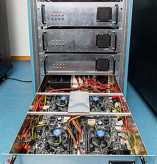
A close-up of one Heinzelmännchen 19" rack drawer, used as a dedicated
cluster for X-ray scanning nano-SAXS data analysis. Using modern EigerX 4M
detectors by Dectris, hundreds of Gigabytes of data can be collected at
Synchrotron Radiation Sources within a few minutes; a parallel analysis of
such large datasets depends on fast analysis hardware and flexible
software, as is currently developed by Markus Osterhoff at the Institute
for X-Ray Physics, Uni Göttingen.
“Using scanning nano-diffraction, we bridge two worlds of X-ray imaging: A
highly focused beam is used to resolve the different constituents inside
biological cells. From the diffraction patterns we get at each position, we
can extract structural information on molecular length scales”, Markus
Osterhoff from Göttingen explains the experimental method. The sample is
raster-scanned and studied at millions of different positions; at each point
in space, the EigerX detector by Dectris produces an “image” with four million
pixels. This can now be done with 750 Hz, leading to data rates of more than
six Gigabytes per second.
Luckily for the researchers and their IT infrastructure, this tremendous data
stream is compressed in real-time. Nevertheless, one Blue Ray disc could be
filled every five to ten minutes. “Recently, we collected one Terabyte of data
per day, for one week of beamtime at the ESRF synchrotron in Grenoble”,
Osterhoff states the problem: within a couple of hours, the number of data
points becomes a 1 with 13 zeros.
Each of the millions of detector images holds more than four million values;
but in the end, every frame is reduced to only one number. Which number? The
group at the university of Göttingen does not yet know. They are establishing
a new quantitative imaging method, and are developing the physical model and
mathematical formulas for that. This means that they have to re-process the
data sets over and over again, changing parameters of the algorithm and
comparing the results.
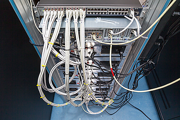
Network connections of the 24-node Heinzelmännchen cluster, used as a
dedicated cluster for X-ray scanning nano-SAXS data analysis.
The group in Göttingen started to analyse the data on specialised hardware they
are otherwise using for X-ray tomography; this three-dimensional technique
needs to handle all images at the same time, and requires large amounts of
memory. For the scanning SAXS method, on the other hand, it turned out that the
actual calculation is very fast, but the amount of data becomes too large for
one many-core parallel computer. Instead, the team acquired 24 consumer-grade
computers, which are now working in parallel. To cut down costs and reduce the
required space, the computers only consist of essential hardware: mainboard,
processor and memory; four nodes share one power supply. ”A control PC
distributes the work and data streams to the computing nodes; now we have 24
`Heinzelmännchen' working on our EigerX datasets, and in principle we could
cope in real-time with four EigerX detectors running in parallel at full
speed”, Markus Osterhoff is proud of their achievement. Now they are combining
the scanning technique with X-ray holography and super-resolution optical
microscopy to get an even sharper and more detailed look into single cells.